
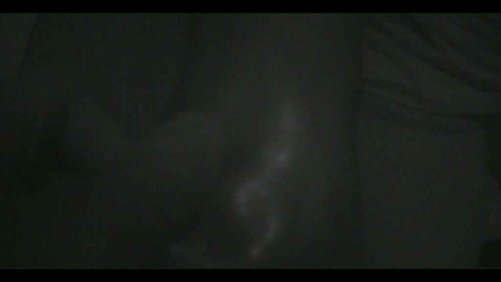
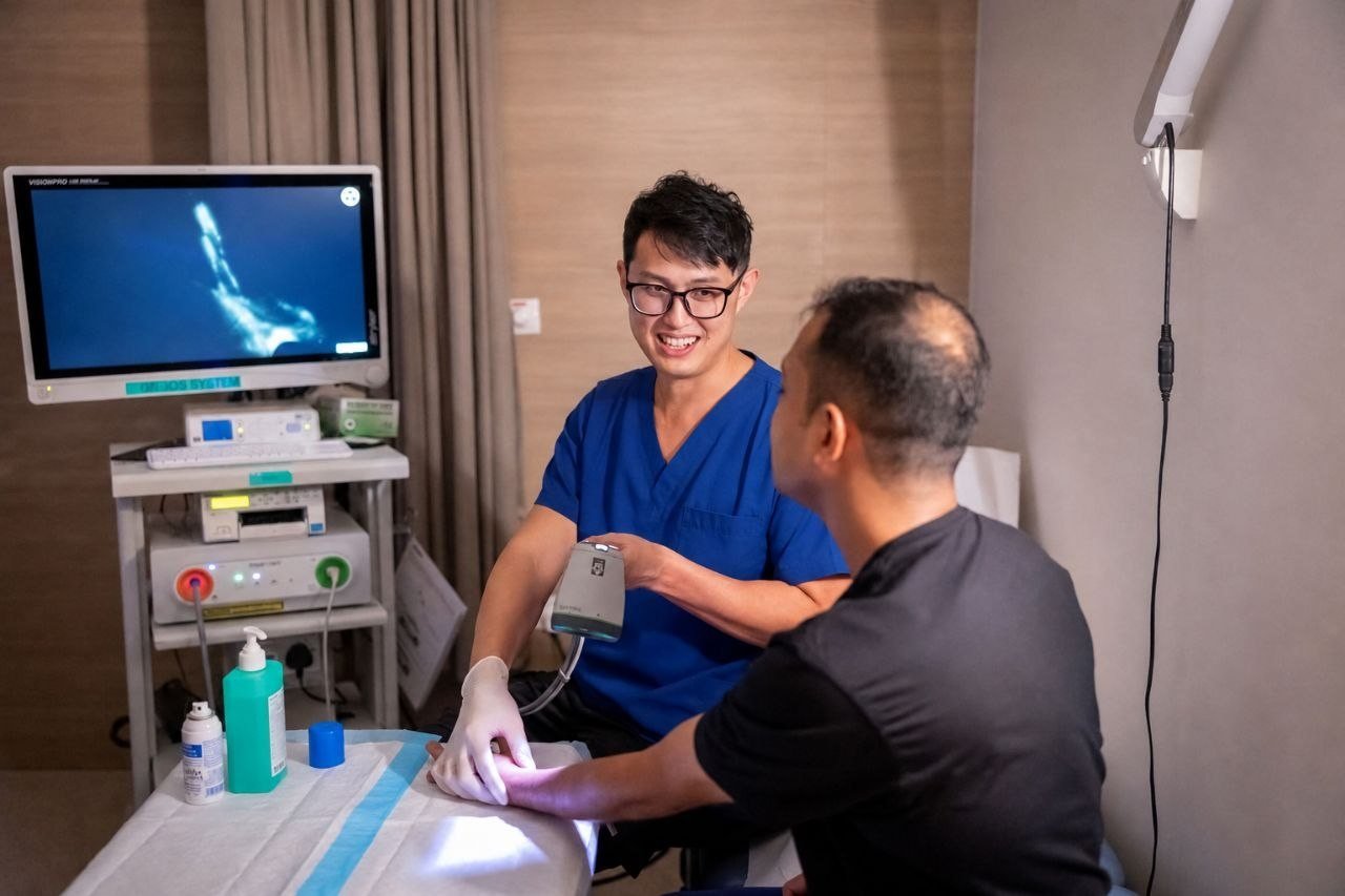
ICG lymphography is a specialised imaging test used to map superficial lymphatic flow in selected patients with suspected or established lymphedema. In Singapore, it may be used as part of specialist assessment when the diagnosis, severity, lymphatic channels, or suitability for microsurgical options such as LVA surgery needs to be clarified.
Specialist-reviewed educational resource: Lymphedema Asia is clinically reviewed by Dr Jeremy Sun Mingfa, a Singapore plastic surgeon with dedicated lymphatic surgery training. This page is educational and does not replace personalised medical consultation.
What is ICG lymphography?
ICG lymphography uses a fluorescent dye called indocyanine green. After small injections into the skin, a near-infrared camera can visualise superficial lymphatic drainage patterns. The test can show whether lymphatic channels are still linear and functional, whether there is dermal backflow, and how lymphatic flow changes across different regions of a limb.
Why it matters in lymphedema assessment
Clinical examination remains important, but imaging can add information that is difficult to judge from swelling alone. ICG lymphography may help with:
- confirming lymphatic dysfunction in early or borderline cases;
- mapping lymphatic flow patterns and areas of dermal backflow;
- planning microsurgical procedures such as LVA surgery in Singapore;
- identifying whether there are potentially usable lymphatic channels;
- monitoring progression or response when clinically appropriate.
ICG lymphography and LVA surgery
For lymphaticovenous anastomosis or lymphovenous bypass, surgeons need to identify suitable lymphatic channels and nearby venous targets. ICG lymphography can help map superficial lymphatics, while ultrasound localisation may also be used in selected cases. Imaging does not assure that LVA is suitable, but it can support a more informed surgical discussion.
What patterns may be seen?
Reports may describe linear lymphatic channels, splash or stardust-like dermal backflow, diffuse patterns, or areas where lymphatic flow is not clearly visualised. These descriptions need to be interpreted in context with symptoms, examination, limb measurements, cellulitis history, prior cancer treatment, and other imaging when relevant.
Who might benefit from specialist assessment?
Specialist review may be helpful for patients with persistent limb swelling, swelling after cancer surgery or radiotherapy, recurrent cellulitis, progressive heaviness or tightness, suspected primary lymphedema, or uncertainty between lymphedema, venous swelling, and lipedema. Related pages include lipedema vs lymphedema, lymphedema treatment and surgery cost factors, and private lymphedema consultation pathways in Singapore.
How ICG findings usually fit into the treatment decision
ICG lymphography is not a stand-alone “yes or no” surgery test. In a specialist assessment, the result is usually interpreted together with the history of swelling, cellulitis episodes, compression response, limb measurements, ultrasound mapping and the patient’s goals.
- Linear lymphatic channels may suggest potentially usable pathways for lymphovenous bypass / LVA surgery, but final suitability still depends on ultrasound and operative findings.
- Dermal backflow or advanced patterns may explain why compression alone is not controlling symptoms, but they do not automatically rule in or rule out surgery.
- Fat- or fibrosis-dominant swelling may require discussion of other options such as lymphedema liposuction or debulking surgery, often with ongoing compression.
- Recurrent cellulitis is a reason to seek careful assessment because infection can further damage lymphatic function. Read more about lymphedema and cellulitis warning signs.
For patients comparing surgical pathways, see the staged overview of LVA vs VLNT vs liposuction for lymphedema and the checklist on whether you may be a candidate for LVA surgery. Patients seeking clinician-specific consultation information can also review Dr Jeremy Sun’s page on lymphovenous bypass / LVA surgery in Singapore.
Limitations and safety considerations
ICG lymphography mainly visualises superficial lymphatics and should not be treated as a standalone diagnosis for every cause of swelling. It may not replace venous assessment, blood tests, cross-sectional imaging, or other investigations when these are clinically needed. Suitability, risks, dye precautions, and alternatives should be discussed with the treating clinician.
Related: Ultrasound mapping for LVA surgery explains why high-frequency ultrasound can identify lymphatic targets even when ICG linear patterns are absent in selected patients.
Core guide: Lymphedema treatment in Singapore explains how diagnosis, compression therapy, ICG lymphography, ultrasound mapping, LVA and advanced-stage surgery fit into a complete treatment pathway.
Frequently asked questions
Is ICG lymphography the same as lymphoscintigraphy?
No. Both assess lymphatic function, but they use different imaging methods and answer different practical questions. The most appropriate test depends on the clinical problem and local availability.
Does an abnormal ICG scan mean I need surgery?
Not necessarily. Many patients need conservative treatment, compression, skin care, physiotherapy, weight optimisation, or monitoring. Surgery is considered only after individual assessment of symptoms, imaging, stage, goals, and risk.
Can ICG lymphography show whether LVA will work?
It can help assess whether suitable superficial lymphatic channels may be present, but it cannot assure outcome. LVA results vary and depend on disease stage, anatomy, surgical findings, compression adherence, and other patient-specific factors.
Related guide: Some patients with painful, heavy or disproportionate legs may have lipedema rather than isolated lymphedema or vein disease. Read: Lipedema diagnosis in Singapore.