Secondary Prevention
Unlike primary prevention, secondary prevention involves early detection of disease. Instituting treatment at this early stage of the disease gives rise to better outcomes, satisfaction with smaller types of surgery.
Regular screening & surveillance
The cornerstone of secondary prevention lies in the consistent screening and vigilant monitoring for the onset of the disease, facilitating early intervention as soon as lymphedema manifests. It is well-established that addressing the condition in its initial stages allows for more straightforward treatments involving less extensive surgeries, and generally yields better outcomes.
To screen for lymphedema, several strategies are in place. From a clinical standpoint, doctors typically assess the severity of symptoms and evaluate patients’ quality of life scores. Additionally, routine measurements of the affected limb’s volume are undertaken. In terms of diagnostic procedures, lymphoscintigraphy and magnetic resonance scans have been the traditional choices. However, these methods come with a set of drawbacks: they are costly, time-intensive, and might not be adept at identifying the disease in its nascent stages.
In light of these challenges, indocyanine green (ICG) lymphography has emerged as a more practical alternative. Not only is it cost-effective and swift, but it also excels in detecting lymphedema in its very early stages, making it a preferred choice in many centers for monitoring disease progression in patients.
When to intervene?
Screening protocols and frequencies vary from institution to institution. For example, a 6 monthly assessment of quality of life scores, volume measurements of the limb, and ICG lymphography can be effective in picking out early disease.
ICG lymphography is usually the first to show signs of lymphedema. When this happens, your doctor will initiate decongestive therapy with manual lymphatic drainage and compression techniques. Lymphovenous anastomosis (LVA) will likely be offered as well since that helps to enhance the drainage of lymphedema fluid. Decongestive therapy alone is helpful in controlling the disease but is unlikely to push patients into a compression-free state.
Remember, the earlier the LVA is done, the higher probability that a patient can be free from compression garments!

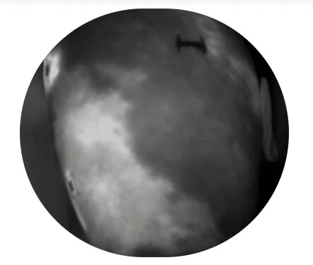
“Splash” and “diffuse” bright spots are typical patterns of lymphedema seen on ICG lymphography.
