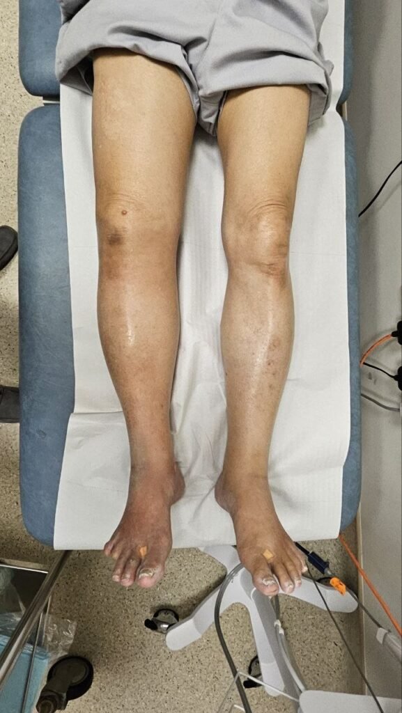
If swelling runs in your family, you have probably spent years wondering whether you carry the same risk. Maybe your mother’s legs were always swollen. Maybe a cousin was diagnosed with something nobody could explain. And now you are watching your own ankles and asking whether what you are seeing is the beginning of the same thing.
Clinically reviewed by Dr Jeremy Sun Mingfa, Senior Consultant Plastic and Reconstructive Surgeon in Singapore, with subspecialty expertise in lymphedema surgery and supermicrosurgical lymphatic reconstruction. Last reviewed: August 2026.
Hereditary lymphedema, including Milroy disease and Meige disease, is real, diagnosable, and better understood now than at any point in history. By the end of this article, you will know the specific genetic forms of hereditary lymphedema, what distinguishes each one, what signs to look for at different stages of life, and what role genetic testing plays in getting an accurate diagnosis.
What Is Hereditary Lymphedema, and How Is It Different from Other Types?
Most lymphedema in the world is secondary, meaning it develops after an event that damages the lymphatic system, such as cancer surgery, radiation therapy, or a parasitic infection. Hereditary lymphedema is different. It is a primary condition, meaning the lymphatic system itself did not form or function correctly from the beginning, because of an inherited gene change.
When a gene that controls lymphatic development carries a fault, the lymphatic vessels may be absent, underdeveloped, or structurally abnormal. The result is that fluid cannot drain properly from the affected area, usually the legs, and it accumulates in the tissue instead. This causes the persistent swelling that defines lymphedema.
Hereditary lymphedema follows an autosomal dominant inheritance pattern in most cases. This means only one copy of the altered gene is enough to cause the condition. If one parent carries the gene change, each child has a 50% chance of inheriting it. However, carrying the gene does not guarantee that swelling will appear. The severity, timing, and even whether symptoms appear at all can vary significantly within the same family, a phenomenon known as variable expressivity.
The clinical term for this family of conditions is primary lymphedema. Historically, doctors classified primary lymphedema by age of onset: congenital (present at birth), praecox (onset between birth and age 35), and tarda (onset after 35). Genetic discoveries have since given a more precise picture, identifying specific genes behind specific subtypes. Named conditions like Milroy disease and Meige disease sit within this genetic framework.
According to the National Organization for Rare Disorders (NORD), primary lymphedema affects approximately 1.15 in 100,000 individuals under 20 years old. It occurs more commonly in females than males, with a ratio of around 3.5 to 1.
What Is Milroy Disease, and What Does It Look Like at Birth?
Milroy disease, also known as hereditary lymphedema type I, is the form of hereditary lymphedema most likely to be visible at birth or within the first year of life. It has been recognized as a distinct clinical entity for over a century, and it is now understood at the genetic level in considerable detail.
What gene causes Milroy disease?
Milroy disease is caused by a fault in the FLT4 gene, which encodes a protein called vascular endothelial growth factor receptor 3 (VEGFR3). According to GeneReviews at the National Institutes of Health, this protein plays a critical role in the development of the lymphatic system during the fetal period. When the FLT4 gene carries a pathogenic variant, the receptor does not function correctly, and the initial lymphatic capillaries, the finest branches of the lymphatic tree, fail to form normally.
The inheritance is autosomal dominant: one altered copy of the gene, inherited from one parent, is sufficient to cause the condition. However, some people carry the FLT4 variant and show little or no swelling, which can make tracking the condition through a family tree more complicated than you might expect.
What does Milroy disease look like clinically?
The defining feature of Milroy disease is lower-limb lymphedema present at or shortly after birth. The swelling typically affects both legs, though it can be asymmetric in some individuals. Parents often notice that a newborn’s feet or lower legs appear puffy in the first days or weeks of life.
Certain physical signs, while not universal, are considered characteristic enough to raise a strong clinical suspicion. These include upward-curling toenails, prominent leg veins that are visible through the skin, deep skin creases on the toes, and small wart-like skin growths called papillomas. Males with Milroy disease sometimes develop a hydrocele, a collection of fluid around the testes, which can also be present at birth.
Recurrent skin infections called cellulitis are a significant complication to be aware of. The stagnant, protein-rich lymph fluid provides an environment where bacteria can thrive, and even a minor skin break can trigger a spreading infection that requires urgent antibiotic treatment. Repeated episodes of cellulitis can cause further damage to the already-compromised lymphatic vessels, worsening the swelling over time.
In clinical practice, this means a child born with persistent leg swelling, particularly if accompanied by any of the characteristic toenail or skin changes, and with a family history of similar swelling, warrants early referral to a specialist with experience in primary lymphedema. Early compression and skin care can meaningfully reduce the risk of cellulitis and slow disease progression.
What Is Meige Disease, and Why Does It Often Appear in the Teen Years?
Meige disease, also called hereditary lymphedema type II or lymphedema praecox, is considered the most common form of primary lymphedema. Unlike Milroy disease, which is evident from birth, Meige disease typically appears around puberty or in the years immediately after, which means many families go through a child’s early years without suspecting anything is wrong, only to see swelling emerge in adolescence.
What triggers the swelling at puberty?
The exact reason puberty acts as a trigger in Meige disease is not fully established. The current understanding is that hormonal changes during adolescence may interact with a lymphatic system that is already structurally borderline, pushing it past the point where it can compensate adequately. The swelling tends to start in one foot or ankle, then gradually progresses up the leg over months or years. A second leg may become affected later.
According to ScienceDirect’s clinical overview of Meige disease, most patients are female, with a female-to-male ratio of approximately 4 to 1, and the swelling in its early stage is typically pitting, meaning it leaves a temporary dent when pressed. Over time, without management, the tissue can become firmer and more difficult to treat.
What gene causes Meige’s disease?
The genetics of Meige disease are less clear-cut than those of Milroy disease. According to the National Organization for Rare Disorders, Meige disease is associated with missense variants in the GJC2 gene in a number of affected families. However, the picture is more complex: genetic testing does not identify a clear causative variant in all patients who have the classic Meige presentation, which means current testing cannot confirm or rule out the condition in every case.
This is an active area of research. The understanding is that Meige disease is likely genetically heterogeneous, meaning more than one gene may be capable of producing the same clinical picture. In clinical practice, this means a negative genetic test result does not automatically rule out hereditary lymphedema in someone whose clinical presentation and family history are consistent with it.
| DR. SUN’S CLINICAL PERSPECTIVE“Patients with Meige disease often tell me they were dismissed for years because their swelling came and went, especially early on. They were told it was hormonal, or that they needed to lose weight, or that nothing could be done.”This means for patients that if you have swelling that started around puberty and a family history of similar symptoms, getting a proper assessment from a specialist with experience in primary lymphedema is worth the effort, even if you have been turned away before. An accurate diagnosis changes the management approach significantly. |
How Do the Three Main Types of Hereditary Lymphedema Compare?
Milroy disease and Meige disease are the most well-known forms of hereditary lymphedema, but they are not the only ones. Lymphedema-distichiasis syndrome is a third distinct genetic form, caused by a different gene and carrying a notable distinguishing feature that can make diagnosis easier when it is recognized. The table below sets out the key differences.
| Milroy Disease | Meige Disease | Lymphedema-Distichiasis | |
|---|---|---|---|
| Also called | Hereditary lymphedema type I / congenital lymphedema | Hereditary lymphedema type II / lymphedema praecox | LD syndrome |
| Gene | FLT4 (VEGFR3) | GJC2 (most cases); genetics not fully established | FOXC2 |
| Inheritance | Autosomal dominant | Autosomal dominant | Autosomal dominant |
| Typical onset | At birth or within the first year of life | Around puberty, up to age 35 | Around puberty or early adulthood |
| Area affected | Lower limbs, usually both legs | Lower limbs, often one side first | Lower limbs; sometimes arms or face |
| Distinguishing feature | Upward-curling toenails, prominent leg veins | Often no other features outside the swelling itself | Double row of eyelashes (distichiasis) – a key clue |
The most striking difference in practice is the eyelash finding in lymphedema-distichiasis syndrome. A second row of eyelashes, growing from the inner part of the eyelid, is called distichiasis. These extra lashes often cause eye irritation, sensitivity to light, and corneal scratching, which sometimes leads the patient to an eye specialist before a lymphedema specialist. According to data cited in OMIM (Online Mendelian Inheritance in Man), approximately 80% of individuals with lymphedema-distichiasis syndrome develop lymphedema by early adulthood.
All three forms are autosomal dominant, meaning they can pass from parent to child regardless of sex. A parent with any of these conditions does not need to have both copies of the gene affected. One copy is enough. This is why families affected by any of these conditions need to understand the inheritance pattern, rather than assuming the condition will skip a generation or only affect one sex.
What Are the Signs That Your Family’s Swelling Could Be Hereditary?
Identifying a hereditary pattern in your family is often the first step toward getting an accurate diagnosis. Not all family members will be equally affected, and some carriers may show only very mild swelling that never gets investigated. Here are the signs that point toward a hereditary cause rather than an acquired one.
- Multiple relatives affected. Swelling that has appeared in more than one family member across different generations, such as a parent and a grandparent, or a parent and a sibling.
- Early onset without a clear trigger. Swelling that started in childhood, adolescence, or early adulthood without any surgery, cancer treatment, injury, or infection that could explain it.
- Lower-limb predominance. Swelling that affects the lower legs and feet specifically, rather than appearing after a clearly identifiable event.
- Associated physical features. Physical signs, such as unusual toenail changes, prominent leg veins, or a double row of eyelashes in some family members.
- Prior unexplained diagnoses in the family. A family member who was previously told they had unexplained chronic swelling, poor circulation, or lymphatic problems without a clear secondary cause.
None of these signs alone confirms a diagnosis. But a combination of two or more, particularly if they span multiple generations, strongly supports a referral for specialist evaluation and genetic testing.
What Does Genetic Testing for Hereditary Lymphedema Actually Involve?
The idea of genetic testing can feel daunting if you have never been through it before. In practice, for hereditary lymphedema, it is a blood test followed by laboratory analysis of specific genes. The process has become considerably more accessible and affordable in recent years as gene panel testing has become standard.
How is the testing done?
Genetic testing for lymphedema typically involves a blood sample or cheek swab, which is sent to a laboratory for DNA analysis. The laboratory looks for changes in genes known to be linked to primary lymphedema. Panel tests, which screen multiple genes at once using next-generation sequencing, are now the standard approach in most specialist settings. A panel targeting lymphedema-related genes can simultaneously check FLT4, FOXC2, GJC2, and other candidate genes in a single test run.
According to a clinical study published in Scientific Reports in 2022 using a targeted panel of 60 genes in patients with primary lymphedema, identifying a confirmed pathogenic variant in all patients remains challenging, reflecting the reality that the genetics of primary lymphedema are still being fully mapped. A positive result confirms a genetic cause and identifies which form you have. A negative result does not rule out hereditary lymphedema, because not all causative variants have been discovered yet.
What information does a genetic test give you?
A confirmed genetic result does several useful things beyond simply giving a diagnosis a name. It establishes the specific subtype of hereditary lymphedema, which can guide treatment decisions and help anticipate how the condition may progress. It also enables your family members to be tested with a targeted approach, looking specifically for the same variant that was found in you, rather than needing a full panel screen.
For people planning families, knowing the inheritance pattern and the specific variant provides accurate information about the risk for future children. This information can be discussed with a genetic counsellor, who helps interpret results in the context of your family situation and your reproductive decisions.
Who should be referred for genetic testing?
Not every patient with swelling needs genetic testing. Testing is most useful in people who have primary lymphedema with onset before age 35, a family history of lymphedema in one or more relatives, physical signs suggesting a known genetic syndrome, such as the eyelash finding in lymphedema-distichiasis, or children and young adults who present with swelling without any identifiable secondary cause.
Testing is typically ordered through a specialist, either a lymphedema specialist or a clinical geneticist, rather than through a general practitioner. The interpretation of results requires clinical context, which is why specialist involvement matters both at the point of ordering and at the point of receiving results.
What happens if no genetic cause is found?
A significant proportion of patients with what appears to be hereditary primary lymphedema will receive an inconclusive or negative result on current gene panels. This is not a failure of the testing process. It reflects the reality that the genetic architecture of primary lymphedema is still being mapped, and researchers expect that additional causative genes remain to be discovered.
In this situation, the clinical diagnosis still stands if the presentation and family history are consistent with hereditary lymphedema. Management proceeds based on clinical assessment rather than waiting for a genetic result. A specialist will typically continue to monitor for new gene discoveries and may recommend retesting as panel technology and gene knowledge evolve. The value of that ongoing relationship with a specialist, rather than a one-time genetic test, should not be underestimated.
Can Hereditary Lymphedema Be Treated, and Does Knowing the Gene Change That?
A common concern among patients newly diagnosed with hereditary lymphedema is whether having a genetic cause means the condition cannot be treated. This concern is understandable but largely unfounded. Treatment for hereditary lymphedema follows the same core principles as treatment for acquired lymphedema, with some important nuances informed by the specific genetic form.
The foundation of management is complete decongestive therapy (CDT), which combines manual lymphatic drainage, a specialized gentle massage technique, with compression bandaging or garments, skin care, and exercise. This approach remains effective regardless of whether the underlying cause is genetic or acquired. The goal is to reduce swelling, maintain skin health, prevent infection, and preserve limb function over the long term.
Early and consistent management produces better long-term outcomes than delaying treatment until complications arise. In clinical practice, this means that children and young adults with confirmed or suspected hereditary lymphedema should start working with a lymphedema therapist promptly, even if the swelling is currently mild. The tissue changes that make lymphedema harder to manage, such as skin thickening and fibrosis, develop over years of inadequately managed fluid accumulation.
For eligible patients, surgical options including lymphovenous bypass (LVB) and vascularized lymph node transfer (VLNT) are also available. These microsurgical procedures aim to create new drainage pathways or introduce functional lymph node tissue into the affected area. The evidence base for surgical management of primary lymphedema is growing, and specialist centres are increasingly evaluating hereditary lymphedema patients for surgical candidacy alongside conservative management.
Knowing the specific genetic form does add some clinical precision. For example, patients with lymphedema-distichiasis syndrome need concurrent management of the eye findings, often with an ophthalmologist, as the extra eyelashes can cause significant corneal damage if untreated. Patients with Milroy disease involving the male genital tract may need evaluation for scrotal lymphedema. These additional considerations make the genetic diagnosis useful beyond just labelling the condition.
What Should You Do If You Think Hereditary Lymphedema Runs in Your Family?
If the information in this article has matched something you have observed in your own family, the most practical next step is a structured clinical evaluation rather than trying to navigate genetic testing independently. Genetic testing for hereditary lymphedema, Milroy disease, and other primary forms works best when ordered and interpreted by a specialist with clinical experience in primary lymphedema.
- Document your family history carefully. Write down which family members have had unexplained swelling, at what age it started, and which part of the body was affected.
- Note any associated physical features. Note any associated features such as eyelash changes, toenail changes, prominent veins, or recurrent skin infections in yourself or relatives.
- Bring photographs of affected relatives. Bring photographs to your appointment if possible, particularly of older relatives who are no longer available for examination, since physical findings can confirm or support a suspected diagnosis even in retrospect.
- Ask about genetic referral. Ask specifically about genetic testing and whether a referral to a clinical geneticist or genetic counsellor is appropriate alongside your lymphedema specialist.
The most important thing to understand is that a hereditary diagnosis does not mean a condition is untreatable or inevitable. It means you have information that most patients with lymphedema never get: a clear explanation for why the condition occurred, and a framework for understanding the risk to other family members. That information is genuinely useful, both for your own care and for the people in your family who may be watching their own legs and asking the same questions you were asking.
Hereditary Lymphedema and Milroy Disease: When to Seek Specialist Input
If you have concerns about hereditary lymphedema or Milroy disease and whether it runs in your family, speaking with a lymphedema specialist early can change your outcome. Dr Jeremy Sun consults at Lymphedema Asia in Singapore, offering specialist assessment, genetic workup coordination, and treatment planning for patients with primary and hereditary lymphedema, including those travelling from overseas for specialist care.