

Your leg has been swollen for weeks. Maybe months. You’ve been told to “elevate it” and “drink more water.” But the swelling isn’t going away, and you’re starting to wonder if something is seriously wrong.
Clinically reviewed by Dr Jeremy Sun Mingfa, Senior Consultant Plastic and Reconstructive Surgeon in Singapore, with subspecialty expertise in lymphedema surgery and supermicrosurgical lymphatic reconstruction. Last reviewed: August 2026.
The lymphatic system helps maintain fluid balance and supports immune function. When lymphatic drainage is impaired, persistent swelling can develop, but swelling also has other possible causes that need appropriate assessment.
By the end of this article, you will understand exactly how the lymphatic system function affects lymphedema, why lymphedema develops, and when it is time to stop waiting and get a proper diagnosis.
What Does Your Lymphatic System Actually Do?
Think of your cardiovascular system as your body’s main highway. Blood travels out from the heart, delivers oxygen and nutrients, and returns. But that highway has a leak problem: every day, about three litres of fluid seep out of your blood vessels into surrounding tissue.
Your lymphatic system is a drainage network that collects this fluid, called lymph, and returns it to the bloodstream. When drainage is impaired, fluid and proteins can accumulate in the tissues and contribute to swelling and tissue changes.
Here is what the lymphatic system does every single day:
- Collects excess fluid from tissues through tiny vessels called lymphatic capillaries
- Filters that filter fluid through lymph nodes, trapping bacteria, waste, and abnormal cells
- Returns clean fluid into circulation via the thoracic duct near your collarbone
- Transports immune cells (lymphocytes) throughout your body to fight infection
The lymphatic system contributes to both fluid balance and immune function. Lymphedema is therefore more than visible swelling, but its effects and severity vary between people.
How Does Lymph Fluid Actually Move Through Your Body?
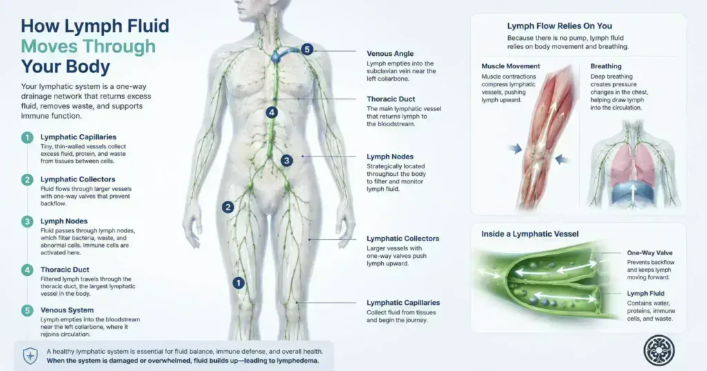
Unlike your heart, your lymphatic system has no pump. It relies on movement, muscle contractions, and breathing to push lymph fluid through a network of vessels that spans your entire body.
Lymphatic capillaries are the starting point. These ultra-thin vessels are present in almost every tissue in your body. Their walls are slightly permeable, which allows excess fluid and protein molecules to pass in from the surrounding tissue.
From there, fluid moves through progressively larger vessels called lymphatic collectors, then into regional lymph nodes. These nodes, you have between 500 and 700 of them, act as checkpoints. They filter the fluid, flag infections, and pass clean lymph forward.
The filtered lymph then flows into the largest lymphatic vessel in your body, the thoracic duct, and empties into the venous system near your left collarbone. Fluid from your right arm and the right side of the head drains through a smaller right lymphatic duct.
In clinical practice, this means that any disruption along this pathway, damaged capillaries, scarred nodes, or blocked collectors, can cause fluid to back up. That backup is what you see as swelling.
Persistent or unexplained swelling needs clinical assessment because lymphedema is one of several possible causes. The lymphedema diagnosis guide explains how history, examination and selected imaging may contribute to that assessment.
What Goes Wrong When Lymphedema Develops?
Lymphedema occurs when the lymphatic transport capacity falls below the volume of fluid the system needs to move. This is not a sudden event; it is typically a slow, progressive failure that begins invisibly.
There are two primary types. Primary lymphedema results from a developmental abnormality in the lymphatic vessels themselves; the system was never fully formed. Secondary lymphedema, the more common form, occurs when a previously healthy system is damaged.
The most common causes of secondary lymphedema include:
- Cancer surgery or radiation that removes or damages lymph nodes (most frequently after breast cancer, melanoma, or gynaecological cancers)
- Infection, particularly filariasis (parasitic infection), is the leading cause of lymphedema worldwide
- Trauma, chronic venous insufficiency, or obesity, placing excessive load on the lymphatic system
When lymph fluid cannot drain, protein-rich fluid accumulates in the interstitial tissue. Over time, this triggers a chronic inflammatory response. Fatty tissue deposits. Skin thickens. The condition progresses through four clinical stages, defined by the International Society of Lymphology (ISL), from reversible early swelling to irreversible hardening of tissue.
Understanding the stage and tissue changes can help guide conservative care, investigations and discussion of whether any procedure is appropriate.
How Is a Healthy Lymphatic System Different from One with Lymphedema?
The table below shows what changes, and why it matters, when lymphedema develops:
| Healthy Lymphatic System | When Lymphedema Develops | |
| Fluid transport | Lymph vessels carry fluid away efficiently | Damaged vessels cannot drain fluid properly |
| Node function | Nodes filter waste and pathogens normally | Nodes may be absent, scarred, or blocked |
| Limb appearance | Normal size and skin texture | Visible swelling, skin changes over time |
| Protein levels | Protein stays in circulation | Protein accumulates in the tissue, worsening swelling |
| Reversibility | A healthy system self-regulates | Requires active treatment; damage can be permanent |
Lymphedema can change over time, and later stages may involve firmer tissue and skin changes. The pattern and pace vary, so persistent, progressive or unexplained swelling should be assessed rather than self-diagnosed from a comparison table.
How Do You Know If Your Swelling Is Lymphedema?
Swelling has many causes, including heart failure, kidney disease, venous insufficiency, and medication side effects are all possibilities. But lymphedema has several distinguishing features that should prompt specialist evaluation.
Pitting vs. non-pitting oedema is one of the first distinctions. In early lymphedema, pressing your finger into the swollen area may leave an indentation (pitting). As the condition progresses and protein accumulates in the tissue, the swelling becomes firmer, and the pit disappears.
Other features that suggest lymphedema over other causes of swelling:
- Swelling that is worse at the end of the day or after prolonged standing, but does not fully resolve overnight
- A feeling of heaviness, tightness, or reduced flexibility in the affected limb
- Skin changes, thickening, a rough texture, or, in later stages, a condition called hyperkeratosis
- Stemmer’s sign: inability to pinch and lift the skin at the base of the second toe (a reliable early clinical indicator)
- History of cancer treatment, infection, or trauma in the affected region
According to ISL consensus guidelines, a clinical diagnosis of lymphedema requires a thorough history, physical examination, and, in many cases, specialised imaging such as lymphoscintigraphy or indocyanine green (ICG) lymphography to assess vessel function directly.
Is It Time to See a Lymphedema Specialist?
Persistent, progressive, asymmetric or otherwise unexplained swelling is a reason to seek clinical assessment. Urgent medical review may be needed when swelling is sudden, painful, red, hot, associated with fever, chest pain or shortness of breath.
You should seek a specialist assessment if any of the following apply:
- Your swelling is persistent, progressive or unexplained
- You have had surgery, radiation, or infection in the affected region, even years ago
- Your swelling is asymmetric: one limb is noticeably larger than the other
Dr Jeremy Sun is a lymphedema microsurgery specialist based in Singapore with clinical expertise in lymphatic system function, lymphedema diagnosis, and surgical reconstruction. His practice, LymphEDasia, serves both local patients and international patients seeking specialist care in Asia.
A lymphedema assessment in Singapore can help distinguish lymphedema from other causes of swelling and guide an individual management plan.
For non-urgent consultation planning, review the private lymphedema consultation pathway.
| KEY TAKEAWAYS |
| ✓ The lymphatic system drains 3+ litres of fluid from your tissues daily and is essential for immune function. |
| ✓ Lymphedema occurs when lymphatic transport capacity fails, typically after cancer treatment, infection, or trauma. |
| ✓ Protein-rich fluid accumulation drives progressive tissue damage through four ISL-defined stages. |
| ✓ Key signs include asymmetric swelling, heaviness, skin changes, and a positive Stemmer’s sign. |
| ✓ Assessment can help clarify the cause of persistent swelling and guide appropriate management. |